|
Continued from The Bird Digestive System
The Intestine
The
intestine,
or gut proper, begins at
the pyloric end of the stomach and ends at the cloaca. It may be conveniently
divided into
-
The duodenum, or first loop.
-
The ileum or narrowest and longest portion,
equivalent to both the jejunum and ileum of humans.
-
The rectum, corresponding with the human large
intestine.
The transition from the ileum to the rectum is marked
by a more or less circular valve (the "ileo-caecal"), so placed as to permit
its contents to pass into the caeca and rectum, but to hinder their return-their
passage throughout the whole intestine being aided by the peristaltic contractions
of the muscular layers of its walls. An epithelium of cylindrical cells,
forming a colourless, structureless and soft cuticle, lines nearly the
whole of the intestine, and is perforated by numerous small pores, opening
upon their interstices. In many parts these cells form very simple and
sometimes tubular glands ("Lieberkuhn's"), and the greater portion of the
walls is beset with the villi mentioned above. These are very numerous,
and are arranged in various ways-being either uniformly and thickly spread
over the surface, giving it a velvety appearance, or are longer and more
sparingly distributed in lines, which may be straight or zigzag, transverse
or longitudinal. Their arrangement is occasionally characteristic of different
groups of birds; but it varies also in different parts of the gut. As a
rule they are largest and most numerous in the duodenum, but sometimes
in the rectum as well. The structure of these small but important
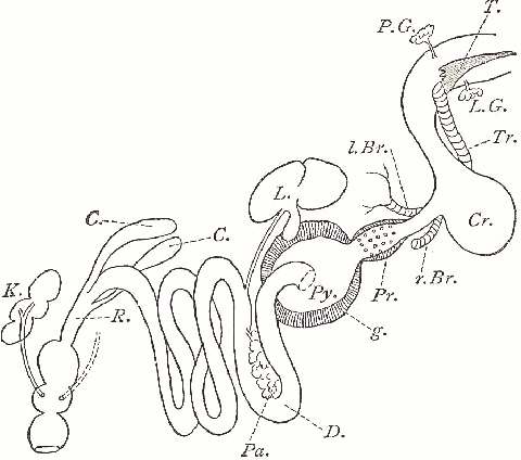
organs will be best understood by reference to the accompanying diagram.
|
Diagram of the Digestive
Organs of a Bird
T. ..... Tongue
P.G. L.G. ..... Parotid and
salivary glands
Tr. ..... Trachea
l.Br. r.Br. ..... left and
right bronchus
Cr. .....Crop
Pr. ..... Proventriculus
or glandular stomach
g. ..... Gizzard or muscular
stomach
Py. ..... Pylorus
D. .....Duodenum
L. ..... Liver with gall-bladder
and duct
Pa. ..... Pancreas with duct
C. ..... Caeca
R. ..... Rectum
K. ..... Kidney with Ureter
opening into the middle cloacal chamber |
The Villi
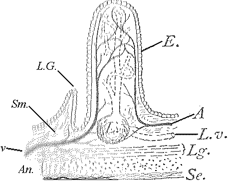
Each villus consists of a finger-shaped prolongation
of the tissue of the submucosa, which contains a ramified central canal
conveying the collected chyle into the lymphatic vessels, which are frequently
connected with a lymphatic follicle for the production of white Blood-corpuscles
or lymph-cells. A pair of small arteries and veins enter the villus, forming
a capillary network, while fine unstriped muscles in its walls contract
it and force the chyle into the lymphatic vessels. In the diagram, on one
side of the villus is shown a Lieberkuhn's gland, since such are generally
associated with the villi.
|
Diagram of an Intestinal
Villus with the Central Absorbent, Ramified Canal
L.v. ..... Its duct
Sm ..... the submucous layer
A and v ..... Artery
and vein ascending in the scubmucous layer
E. ..... Cylindrical cells
of the epithelium of the mucous layer, which at L.G. forms a Lieberkuhn
gland
Lg. and An. ..... Longitudinal
and annular or circular muscular fibres
Se. ..... Serosa or outer
layer of connective tissue, together with the investing peritoneal lamella
Pe., which forms the mesentery
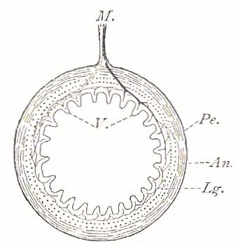
M.
in the diagram below. |
|
Diagram of a Transverse Section
through the Intestine
V. ..... Villi
M. ..... Mesentery with blood
and lymphatic vessels. |
|
